Optical 3D microscopy can enable more effective diagnosis of kidney diseases
A new method developed by KTH researchers for advanced 3D sample preparation and 3D microscopy can make diagnosing kidney diseases more effective.
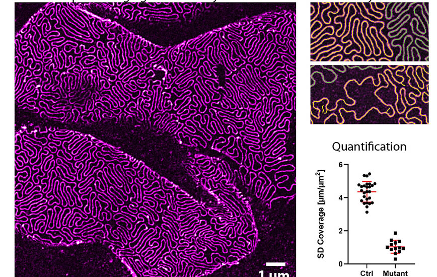
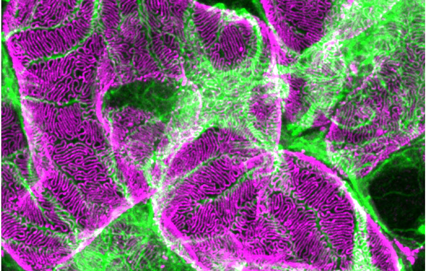
“A single tissue sample provides high resolution image material of the kidney that can be directly analysed in three dimensions,” says Hans Blom, Facility Head at SciLifeLab, the national bioscience hub in Stockholm.
More than half a million people are estimated to live with chronic kidney diseases in Sweden. The problem is exacerbated as people the world over are increasingly developing diseases such as diabetes and high blood pressure, which in turn, affect the way kidneys function. Added to which, COVID-19 has often proven to cause acute damage to kidneys.
“This new 3D microscopy makes current diagnostic processes more effective. Our method is quicker to perform – we can go from a kidney biopsy to ready images in five hours. The corresponding process used today takes 24 hours or more,” says David Unnersjö-Jess, who has developed the new technology together with Docent Hans Blom and Professor Hjalmar Brismar at KTH.

In the existing electron microscopy method, sections of tissue samples are studied, although these sections provide only a fraction of the total information, Blom says.
“This means you can easily miss a kidney diagnosis,” Blom says. “In our new 3D method you can see the whole sample, and it uses simpler equipment, takes less time and requires less manual input. You only need one instrument instead of three, and one protocol instead of four.”
The research group has recently published the protocol in one of the largest periodicals for kidney research , together with clinical kidney researchers, and is now working to validate the protocol for broader clinical use.
Are clinical physicians positive about the new technology?
“It has received a very varied reception, which has ranged across the entire scale from enthusiasm to scepticism in Sweden, Italy and Germany. Many doctors who have dedicated their entire professional lives to the classic diagnosis of renal disease are less willing to have a rethink. However, we view criticism as a sounding board,” Unnersjö-Jess says.

He is further developing the protocol at the NephroLab in Cologne, a medical laboratory in Germany where the majority of researchers have degrees in medicine.
“I was recently invited to use this 3D technology to help a clinic in Cologne when a kidney patient’s electron microscopy images were unable to show any damage. Using 3D microscopy and the analytical tools we have developed, we were able to rapidly see a clear 15-percent change in the small filtration structures of the kidney that electron microscopy had missed,” he says.
What weaknesses does this new 3D technology have compared to the standard diagnostic method?
“One small disadvantage is that we are not able to achieve the same enormous high resolution as in classical electron microscopy, where you can see molecular structures in certain sections. However, the strength we have is that we can keep the tissue studied intact and view the whole of the 3D image quickly and easily,” Blom says.
3D microscopy is currently being evaluated for use within clinical research at the Karolinska Hospital in Huddinge.
When can the technology become an established part of healthcare?
“Clinical use of health technology is conservative and it takes time to train personnel, so we envisage this will be in a couple of years. In the initial phase, we view our new 3D method as a complement above all to existing diagnostic technology,” Unnersjö-Jess says .
Katarina Ahlfort
Photo/illustration: David Unnersjö-Jess